Γλώσσα
 Ελληνικά
Ελληνικά
Ελληνικά
MAGUS
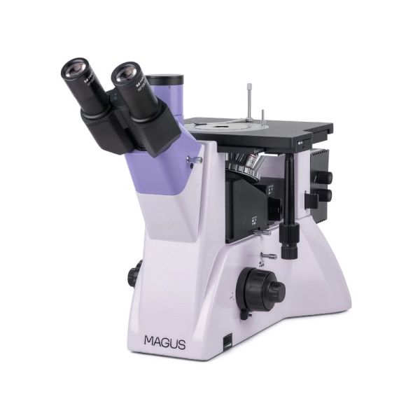
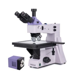
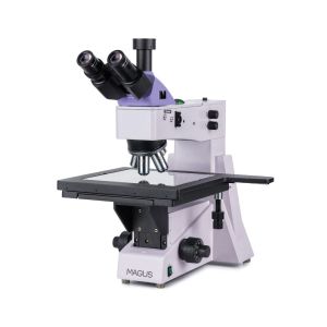
The microscope is designed for studying the microstructure of metals and alloys, semiconductor materials, and other opaque specimens.
Διαθέσιμο κατόπιν παραγγελίας

MAGUS METAL V700
Key features
Microscope head
Tube length – infinity (∞). When assembling the microscope, the user can rotate the eyepiece tubes 180° and adjust the eye relief to fit their height. There is a trinocular head.
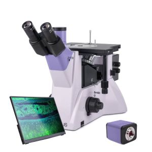
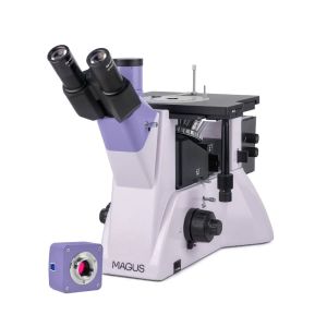
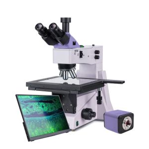
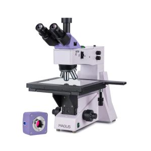
The monitor is mounted in the trinocular tube of the trinocular head and the digital camera – in the camera side port on the microscope body. The trinocular head is equipped with an 50:50 beam splitter, and there is a 2-position beam splitter (100:0/ 0:100) on the body.
Revolving nosepiece
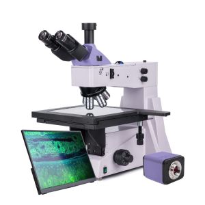
The 5-objectives revolving nosepiece is mounted on the stand under the stage.
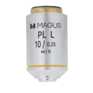
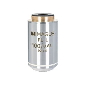
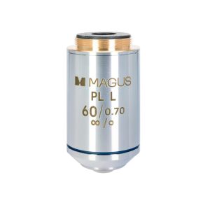
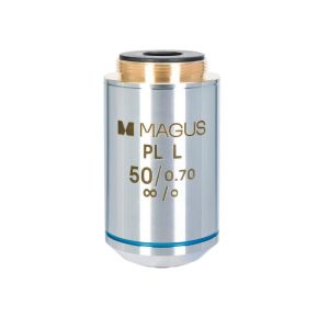
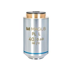
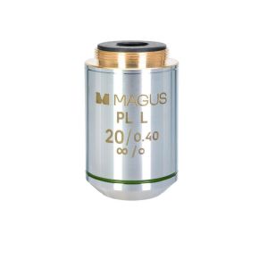
Objectives
Plan achromatic objectives with a long working distance that is designed for the brightfield microscopy technique. For the darkfield microscopy technique, choose the MAGUS Metal V700 BD.
Focusing mechanism
Coaxial coarse and fine focusing knobs are located at the base of the microscope on both sides. The user can place their hands on the table and take a relaxed pose while observing. The focus adjustment is smooth and effortless. On the right, there is the coarse focusing lock knob for quick adjustments after changing the sample. On the left side, there is the coarse focusing tension adjusting ring.
Mechanical stage
The object is moved by moving the stage along two axes. A round rotating stage plate with a hole of suitable diameter (10mm, 20mm, or 30mm) is installed in the center of the table. A specimen holder is used for holding the object in place.
Light source
The lamphouse has a 30W halogen bulb. It is bright enough to observe with 4x to 100x magnification objectives that are used for the brightfield and polarized light microscopy techniques. The halogen bulb emits light with a color temperature that allows for comfortable work.
Reflected light illumination
The illumination system makes it possible to set up the Köhler illumination. The field and aperture diaphragm are pre-centered at the factory and require no additional centering. If necessary, the diaphragms can be adjusted with centering screws. The light source is centered along three axes. The removable analyzer and polarizer are for the polarization microscopy technique. The polarizer rotates 0–360°, the analyzer does not rotate. A set of filters can help you adjust the color reproduction.
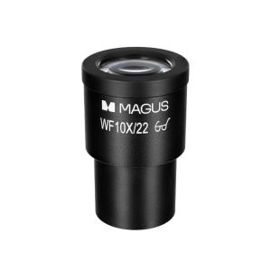
Accessories
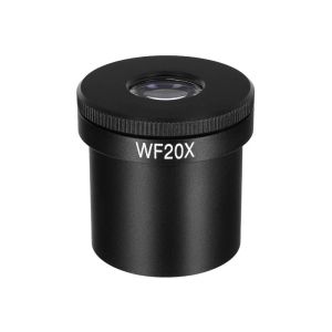
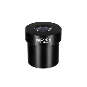
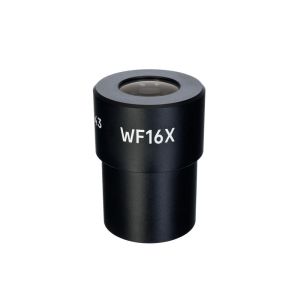
Additional accessories will increase the technical performance of the microscope. Eyepieces and objectives extend the magnification range. Digital cameras output the image to a computer screen or monitor. Calibration slides help you take measurements of objects.
Specifications
In the box:
Available on request:
Όλες οι παραγγελίες που δεχόμαστε μέχρι τις 15:00 και αφορούν προϊόντα που βρίσκονται στην αποθήκη μας, διεκπεραιώνονται την ίδια μέρα
Άρτια εκπαιδευμένο επιστημονικό και τεχνικό προσωπικό, είναι στη διάθεσή σας ανά πάσα στιγμή, για να λύσει τις απορίες και τους προβληματισμούς σας σχετικά με τον εξοπλισμό σας.
Σε περίπτωση που δεν μείνετε ικανοποιημένος/η από το προϊόν που παραλάβατε, μπορείτε να το επιστρέψετε εντός 14 ημερών από την ημερομηνία παραλαβής του.
Εμπειρία που εμπιστεύεστε . Το Πλανητάριο ΘΕΣΣΑΛΟΝΙΚΗΣ είναι ο μεγαλύτερος προμηθευτής ειδών αστρονομίας στα Βαλκάνια
 English