Language
 English
English
English
MAGUS
The camera makes it easy to work with samples that are moving and is suitable for classroom demonstrations. The high-speed camera is recommended for professional laboratories, research, and university training.
Available Upon Request

This includes products that we do not keep in stock but are only available upon request or have been announced in advance by the manufacturer. These items are usually shipped within 5-14 days. Once we receive your order, we will notify you by e-mail or telephone of the exact delivery time, as well as the amount of any deposit that will be required.
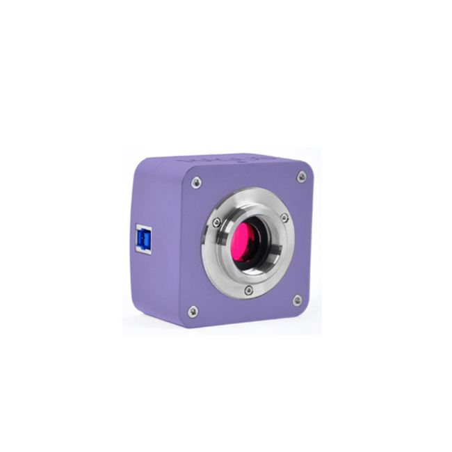
MAGUS CBF90 DIGITAL CAMERA
Features:
Sensor
The camera is equipped with a SONY Exmor backlit color CMOS sensor. This technology increases light sensitivity and improves camera performance in low-light conditions.
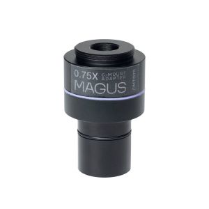
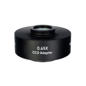
Large sensor – 2/3''. The size of the sensor affects the image quality: The larger the sensor is, the cleaner the image will be, and the greater the color depth and the wider the field of view. The 2/3" sensor with 3.45x3.45µm pixel provides light sensitivity of 1146mV at 1/30s. To achieve the maximum field of view without distortion, we recommend using an adapter with a magnification of 0.7x to 0.9x. It is one of the most efficient cameras for brightfield microscopy with 20x, 40x, 60x, and 100x objectives.
Software features
The camera uses software to display images on an external display, take photographs, record video, edit images, and measure linear and angular dimensions of objects and their structures. Before taking measurements, the software must be calibrated for each objective with a calibration slide.
Installation
The camera is mounted on the microscope in the trinocular tube or side camera port using the C-mount adapter (included). To mount the camera in an eyepiece tube instead of an eyepiece, you will need a special C-mount adapter and adapters to fit the tube diameter (not included).
In the box:

All orders we receive until 15:00 concerning products in our warehouse, processed on the same day

Well-trained scientific and technical personnel are available to you at any time, to solve the questions and concerns about your equipment.

If you are not satisfied with the product you received, you can return it within 14 days of receipt.

Experience you can trust. Planetarium of Thessaloniki is the largest supplier of astronomy goods in the Balkans
 Ελληνικά