Γλώσσα
 Ελληνικά
Ελληνικά
Ελληνικά
MAGUS
The microscope is designed to study objects in polarized and natural light.
In the transmitted light, you can study geological specimens as well as anisotropic biological and polymeric specimens in thin sections.
Διαθέσιμο, συνήθως σε 5-14 μέρες


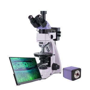
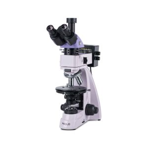
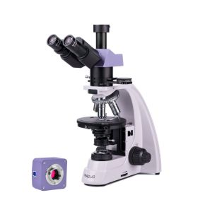
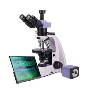
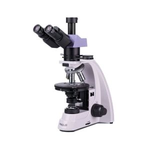
MAGUS POL 850
Key features of the microscope:
Key features of the camera:
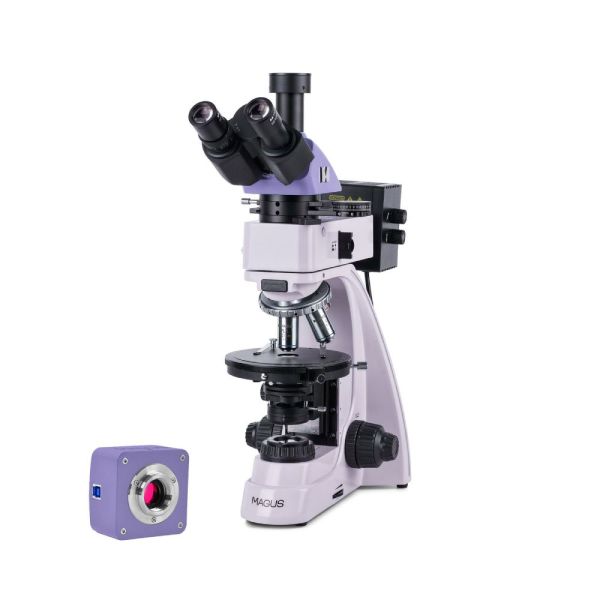
Digital camera
MAGUS CBF70 is a science camera with 21MP resolution and 4/3" sensor for the brightfield microscopy technique.
The camera delivers a realistic image at a 5280x3954 pix resolution. The camera is recommended for use with 4x, 10x, 20x, and 40x objectives. When working with low magnification objectives, the camera will allow you to see finer details.
At maximum resolution, video is captured at 17fps. It is enough for adjusting the focusing of the microscope.
When the resolution is reduced, the rate increases to 56, 67, and 192fps. Transitions between frames become softer, the camera catches the slightest movement of the sample. These modes are suitable for classroom demonstrations, recording video of fast-moving processes, and observing samples that are moving.
The camera is equipped with a USB 3.0 interface. The data transfer speed is 10 times faster than USB 2.0 cameras. The high-speed camera is recommended for professional laboratories, research, or university training.
Microscope head
Trinocular head with infinity plan achromatic objectives. Digital image capturing devices are mounted in the trinocular tube. The eyepiece/camera light path selection knob allows you to direct the light beam to either the digital camera or the eyepiece tubes.
The diopter adjustment is on the left tube.
Revolving nosepiece
Five objectives. A free slot is used for centering the light source. An additional objective can also be installed in a free slot in order to achieve extra magnification. The design of the revolving nosepiece (“away from the observer”) frees up space at the front of the stage and, therefore, the user can see the objective inserted into the optical path. The revolving nosepiece slots are centered to align the optical axis of the objective and microscope.
Objectives
Infinity plan achromatic objectives are designed specifically for polarized light observations: The strain-free optics ensure that the birefringence comes from the specimen and not from the optical elements. The objectives are designed to study samples without cover slips.
Focusing mechanism
Coaxial coarse and fine focusing knobs are located at the bottom of the stand on both sides. The user can place their hands on the table and take a relaxed pose while observing. The focusing adjustment is smooth and effortless.
The design of the focusing mechanism provides for quick microscope adjustment after changing the object of study. For this purpose, a coarse focusing lock knob is located on the right side. The coarse tension can be adjusted by turning the ring on the left side.
Stage
The stage rotates 360° to view the color change of the sample when the polarizer and analyzer are in crossed orientation. The stage has a gradation of the rotation angle. With the vernier scale, measurements are made with an accuracy of 0.1°.
The stage can be centered with two screws because the analysis of an anisotropic object in polarized light requires the precise alignment of the rotation axis of the stage with the optical axis of the microscope.
Light source
30W halogen bulbs are used in both reflected and transmitted light illuminators. Halogen bulbs emit light with a color temperature that allows for comfortable work. The 30W bulb is bright enough for you to observe with the 4x to 100x magnification objectives that are used for brightfield and polarized light microscopy.
Reflected light illumination
The illumination system makes it possible to set up the Köhler illumination. The field and aperture diaphragms are pre-centered at the factory and require no additional centering. If necessary, the diaphragms can be adjusted with centering screws. The light source is centered along three axes. The removable analyzer and polarizer are for the polarization microscopy technique. The polarizer rotates 0–360°, the analyzer rotates 360°, and the vernier scale on the analyzer provides accurate angle measurement.
A set of filters can help you adjust the color reproduction.
Transmitted light illumination
Köhler illumination: adjustable field diaphragm; centered and height-adjustable Abbe condenser with adjustable aperture diaphragm and swivel auxiliary lens. NA = 1.25. The polarizer is 0–360° rotatable, with four rotation angles of 0°, 90°, 180°, 270° marks on the scale. The analyzer rotates 360°, the vernier scale on the analyzer provides accurate angle measurement.
Köhler illumination in reflected and transmitted light
The Köhler illumination setup enhances the image quality of a specimen. With such illumination, you can achieve maximum resolution on each objective and uniform illumination of the field of view with no darkening at the edges. The object of study is in sharp focus, and the image artifacts are removed.
Studying samples in polarized light
The intermediate tube holds the analyzer and Bertrand lens, and it has a slot for compensators. The analyzer is inserted into the light path to observe objects in polarized light. When the polarizer and analyzer rotate, the polarization angle changes. You can achieve a mutually perpendicular position by setting both filters to the “0” position. When the microscope stand is rotated, depending on the polarization angle, the surface of the sample reflects light differently. The Bertrand lens is used for conoscopic studies. The compensators are designed to enhance the contrast of samples with weak birefringence.
Accessories
There is a line of accessories designed for the microscope.
Additional objectives can be used for studying objects up to 0.32µm.
Eyepieces extend the magnification range of the microscope. Additional eyepieces will help to utilize the full potential of an objective that is used more often.
A digital camera that outputs the microscope image on a monitor, stores files, and that has software that takes real-time measurements of specimens. A calibration slide for measuring objects that can be combined with an eyepiece with a scale or the camera software.
Microscope specifications
Camera specifications
In the box:
Available on request:
Όλες οι παραγγελίες που δεχόμαστε μέχρι τις 15:00 και αφορούν προϊόντα που βρίσκονται στην αποθήκη μας, διεκπεραιώνονται την ίδια μέρα
Άρτια εκπαιδευμένο επιστημονικό και τεχνικό προσωπικό, είναι στη διάθεσή σας ανά πάσα στιγμή, για να λύσει τις απορίες και τους προβληματισμούς σας σχετικά με τον εξοπλισμό σας.
Σε περίπτωση που δεν μείνετε ικανοποιημένος/η από το προϊόν που παραλάβατε, μπορείτε να το επιστρέψετε εντός 14 ημερών από την ημερομηνία παραλαβής του.
Εμπειρία που εμπιστεύεστε . Το Πλανητάριο ΘΕΣΣΑΛΟΝΙΚΗΣ είναι ο μεγαλύτερος προμηθευτής ειδών αστρονομίας στα Βαλκάνια
 English