MAGUS BIO VD300

MAGUS BIO VD300

MAGUS BIO VD300

MAGUS

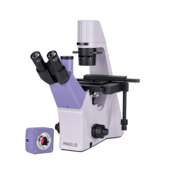
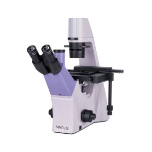
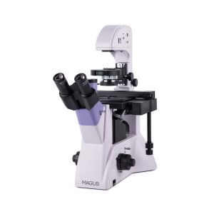
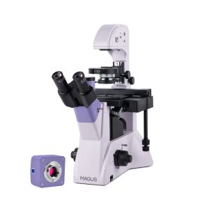
This microscope is designed for studying liquid precipitates, cell colonies, living cells, tissue cultures, and other stained and unstained specimens in laboratory glassware. Its inverted design allows for the use of Petri dishes, multi-well plates, vials, roller bottles, and flasks up to 70mm with a bottom thickness of 1.2mm. 

3.149,00 €

Διαθέσιμο με Προπαραγγελία

Διαθέσιμο με Προπαραγγελία

Στην περίπτωση αυτή περιλαμβάνονται προϊόντα που συνήθως δεν κρατάμε σε στοκ και για κάποιους λόγους - που δεν εξαρτώνται από την εταιρία μας - ΔΕΝ μπορούν να είναι διαθέσιμα σε διάστημα μικρότερο των 14 ημερών.Στην κατηγορία αυτή περιλαμβάνονται και προϊόντα που προδιαφημίζονται από τους κατασκευαστές αλλά δεν άρχισε ακόμη η επίσημη διανομή τους στην ελληνική αγορά. Πιθανόν για τα είδη αυτά να παρέχεται δυνατότητα προπαραγγελίας με ειδική προνομιακή τιμή. Αν γνωρίζουμε ήδη την πιθανή ημερομηνία άφιξης του συγκεκριμένου προϊόντος, στις αποθήκες μας, τότε αυτή θα αναγράφεται ξεχωριστά και επάνω από την ένδειξη «Διαθέσιμο με Προπαραγγελία»

Διαθεσιμότητα: Σε απόθεμα
SKU
LEV-83012


MAGUS BIO VD300


Key features of the microscope:

  • Studies of stained and unstained objects in lab glassware (Petri dishes, flasks, plates)
  • Microscopy techniques: brightfield and phase contrast
  • Trinocular head with a trinocular tube for mounting a digital camera; 50:50 beam splitting
  • Transmitted light illuminator: energy-saving 9W LED with a lifetime of up to 50,000 hours
  • Condenser with a phase contrast slider, phase rings that can be centered
  • The stage is equipped with a mechanical attachment for moving lab glassware along the X and Y axes; dish holders are included in the set
  • Additional accessories


Key features of the camera:
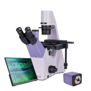
  • 4K camera for brightfield and darkfield microscopy observations with 4x, 10x, and 20x objectives as well as stereomicroscope: The higher the resolution is, the finer the details will be that you can see.
  • 45fps at 4K resolution, 70fps at Full HD resolution: easy focus adjustment, classroom demonstrations, observation of moving samples, and video recording - the movement of the sample can be seen without jerkiness and lags.
  • SONY Exmor backlit color CMOS sensor provides a low noise level and high light sensitivity even in low-light conditions. You will get clearer, brighter, and more color-saturated images.
  • USB 3.0 interface for fast, smooth data transfer
  • Software with photo, video recording, editing, external display, and linear and angular measurements


Digital camera

The MAGUS CDF30 digital camera is designed for the brightfield and darkfield microscopy technique. It offers a low noise level and high light sensitivity.
The camera is equipped with an 8.3MP sensor and delivers realistic images in 4K resolution (3840x2160pix) with the clearly visible fine details of samples and their structures. It is recommended to use the camera with 4x, 10x, and 20x objectives as well as with a stereomicroscope.
At maximum resolution, video is captured at 45fps. This is enough for classroom demonstrations, focus adjustment, recording fast-moving processes, and observing moving samples. At Full HD resolution (1920x1080pix), the rate increases to 70fps, and so the transitions between frames become softer and the camera can catch the slightest movement of the sample.
The camera is equipped with a USB 3.0 interface. The data transfer speed is 10 times faster than USB 2.0 cameras. The high-speed camera is recommended for professional laboratories, research, or university training.


Microscope head

A trinocular head with infinity plan achromatic objectives. The digital camera is mounted in the trinocular tube. 50:50 beam splitting.


Revolving nosepiece

A 4-objective revolving nosepiece that is mounted on a stand under the stage.


Objectives

Infinity plan achromatic objectives with a long working distance for dishes with a bottom thickness of 1.2mm. The four objectives are for the brightfield microscopy technique and one objective – for the phase contrast technique.


Focusing mechanism

The coaxial coarse and fine focusing knobs are located at the base of the microscope on both sides. The user can place their hands on the table and take a relaxed pose while observing. The focus adjustment is smooth and effortless. On the right side, there is a coarse focusing lock knob for quick setting adjustments after changing the sample. On the left side, there is a coarse tension adjustment ring for further adjustment.


Stage

There is a fixed stage with a mechanical attachment for moving the laboratory glassware in two mutually perpendicular directions. The smooth and subtle movement of the object augments the accuracy of the study: No part of the specimen will be overlooked. The travel knob is located at the bottom, and so the user will not strain their right wrist. The microscope set includes three dish holders for glassware of different sizes.


Condenser

A phase contrast slider is installed in the condenser. The phase contrast rings can be centered. The installation of such a slider saves time when switching between microscopy techniques.


Light source

The 9W LED provides bright illumination, which is sufficient for observations in brightfield and phase contrast with all kinds of objectives. The color temperature does not change when you adjust the brightness. The LED has a lifespan of 50,000 hours.


Accessories

There is a line of accessories designed specifically for this microscope. There are additional 20x and 40x phase objectives for studying 0.7µm and 0.4µm non-contrast objects. Eyepieces extend the magnification range of the microscope. Additional eyepieces will help you use the full potential of an objective that is used more often. A digital camera that outputs the microscope image to a monitor and stores files, and the software that takes real-time measurements of specimens. A calibration slide for measuring objects that can be combined with an eyepiece with a scale or the camera software.

Microscope specifications

  • Microscope design: inverted
  • Image field: flat
  • Transmitted light microscopy techniques: brightfield, phase contrast
  • Microscope magnification, x: 100–400 basic configuration (*option: 100–500/600/800/1000)
  • Tube length: infinity (∞)
  • Microscope head type: trinocular
  • Microscope head: Siedentopf
  • Inclination: 45°
  • Interpupillary distance, mm: 48–75
  • Eyepiece diameter, mm: 30
  • Eyepieces, x/field, mm: 10х/22mm, eye relief: 10mm (*optional: 10x/22mm with a scale, 12.5x/14; 15x/15; 20x/12; 25x/9)
  • Revolving nosepiece: 4 objectives
  • Optical design: infinity plan achromatic objectives (∞), parfocal distance: 45mm
  • Objectives, x/aperture/working distance, mm: PL 10x/0.25/4.27; PL 20x/0.40/8.0; PL 40x/0.60/3.5; PHP2 10x/0.25/4.27 phase (*optional: PHP2 20x/0.40/8.0 phase; PHP2 40x/0.60/3.5 phase)
  • Spring-loaded retractable assembly: 40х
  • Stage: fixed, with mechanical attachment for moving the specimen; dish holders: 29x77mm, Ø90mm; 34x77.5mm, Ø68.5mm; 57x82mm, Ø60mm
  • Moving range, mm: 77/112
  • Condenser: NA 0.6, working distance: 70mm; with adjustable aperture diaphragm and a phase contrast slider slot
  • Focusing mechanism: coaxial coarse and fine focusing knobs located on both sides
  • Fine focusing scale value, μm: 2
  • Coarse focusing tension adjusting knob: +
  • Coarse focusing lock knob: +
  • Illumination: transmitted
  • Transmitted light source: 9W LED, brightness-adjustable
  • Phase contrast device: phase contrast slider for 10x objective lens with a phase ring that can be centered
  • Power supply, V/Hz: 220±22V/50Hz via an AC power supply
  • Operating temperature range, °C (°F): 5… 35 (41… 95)
  • Operating humidity range, %: 20… 80


Camera specifications

  • Sensor: SONY Exmor CMOS
  • Color/monochrome: color
  • Megapixels: 8.3
  • Maximum resolution, pix: 3840x2160
  • Sensor size: 1/1.2'' (11.14x6.26mm)
  • Pixel size, μm: 2.9x2.9
  • Light sensitivity: 5970mV/lux/s
  • Signal/noise ratio: 0.13mV at 1/30s
  • Exposure: 0.02ms–15s
  • Video recording: +
  • Frame rate, fps at resolution: 45@3840x2160; 70@1920x1080
  • Place of installation: trinocular tube, eyepiece tube instead of eyepiece
  • Image format: *.jpg, *.bmp, *.png, *.tif
  • Video format: *.wmv, *.avi, *.h264 (Win 8 or newer), *h265 (Win 10 or newer)
  • Spectral range, nm: 380–650 (with IR filter)
  • Shutter type: ERS (electronic rolling shutter)
  • White balance: auto/manual
  • Exposure control: auto/manual
  • Software features: image size, brightness, exposure time
  • Output: USB 3.0, 5Gb/s
  • System requirements: Windows XP (32 bit), Vista/7/8/10/11 (32 and 64 bit), Mac OS X, Linux, up to 2.8GHz Intel Core 2 or higher, minimum 2GB RAM, USB 3.0 port, CD-ROM
  • Software: MAGUS View
  • Mount type: C-mount
  • Body: solid aluminum
  • Power supply: DC current, 5V, with the USB port of the PC
  • Operating temperature range, °C: −10... 50
  • Operating humidity range, %: 30… 80


In the box:

  • MAGUS CDF30 Digital Сamera (digital camera, USB cable, installation CD with drivers and software, user manual and warranty card)
  • Base with a power input, transmitted light source, focusing mechanism, condenser, and revolving nosepiece
  • Trinocular head
  • Infinity plan achromatic objective: PL 10x/0.25 WD 4.27mm
  • Infinity plan achromatic objective: PL L20х/0.40 WD 8.0mm
  • Infinity plan achromatic objective: PL L40х/0.60 WD: 3.5mm
  • Infinity plan achromatic objective: PHP2 10x/0.25 phase WD 4.27mm
  • Eyepieces 10x/22mm with long eye relief (2 pcs.)
  • Phase contrast slider with centering phase rings
  • Fixed stage
  • Mechanical attachment for moving the specimen
  • Cup holders (3 pcs.)
  • AC power cord
  • Dust cover
  • User manual and warranty card


Available on request:

  • 10x/22mm eyepiece with a scale
  • 12.5x/14mm eyepiece (2 pcs.)
  • 15x/15mm eyepiece (2 pcs.)
  • 20x/12mm eyepiece (2 pcs.)
  • 25x/9mm eyepiece (2 pcs.)
  • Infinity plan achromatic objective: PHP2 20x/0.40 phase WD 8.0mm
  • Infinity plan achromatic objective: PHP2 40x/0.60 phase WD 3.5mm
  • Calibration slide
Γράψτε τη Δική σας Αξιολόγηση
Μόνο οι εγγεγραμένοι χρήστες μπορούν να γράψουν αξιολογήσεις. Παρακαλώ, συνδεθείτε ή δημιουργήστε ένα λογαριασμό
Σχετικά Προϊόντα: