Γλώσσα
 Ελληνικά
Ελληνικά
Ελληνικά
MAGUS
The microscope is suitable for observing transparent and translucent biological samples, such as smears and cross sections using the brightfield microscopy technique in transmitted light. The installation of additional accessories will allow for darkfield, phase-contrast, fluorescence, and polarization methods.
Διαθέσιμο, συνήθως σε 5-14 μέρες


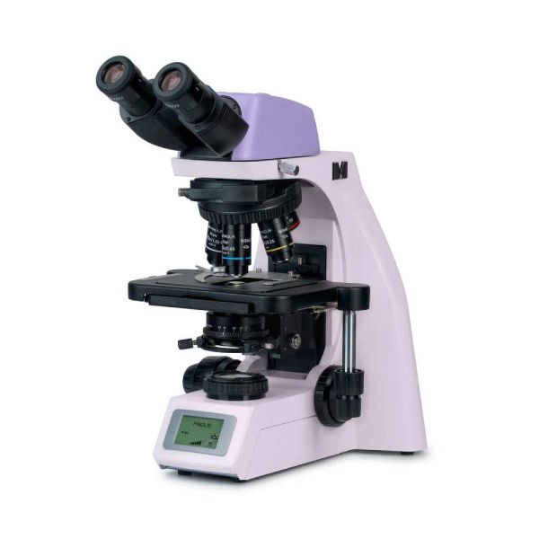
MAGUS Bio DH260 Biological Digital Microscope
Features:
The microscope is suitable for observing transparent and translucent biological samples, such as smears and cross sections using the brightfield microscopy technique in transmitted light. The installation of additional accessories will allow for darkfield, phase-contrast, fluorescence, and polarization methods.
The coded revolving nosepiece maintains a comfortable brightness level when switching objectives. Intelligent control of microscope lighting increases the comfort and speed of the researcher’s routine work. “Smart” functions help students more easily enter the profession and gain the necessary professional experience.
A digital camera is built into the microscope head. The microscope has a minimalist design without a vertical tube or protruding parts.
The microscope is suitable for lab work, research, and teaching.
Microscope head
Binocular head with infinity-corrected optics. Eyepiece tubes rotate 360°. The user can adjust the eye relief on the eyepiece to fit their height.
The base kit includes 10x/22mm eyepieces with diopter adjustment and a long eye relief for working with glasses. Flat rubber eyecups protect the optics from scratches.
Digital camera
A digital camera is built into the microscope head.
The camera is equipped with an 8MP sensor and delivers realistic images in 4K resolution (3840x2160px) with the clearly visible fine details of samples and their structures. The best images are produced using 4x and 10x objectives.
Video is recorded at 30 fps at maximum resolution. Videos are smooth, with soft and subtle transitions between frames. The movement of the sample is displayed in real time with no delays. The camera provides convenient work with moving objects and is suitable for conducting demonstrations in a classroom, photographing samples, and fine-tuning the focus of a microscope.
Wi-Fi is used to connect to a computer and transfer data to it.
Revolving nosepiece and objectives
The coded revolver for 5 objectives is oriented toward the interior: The user can see the objective inserted into the optical path, and the space above the stage is free. An additional objective can also be installed in the free slot in order to achieve extra magnification. The parfocal distance is 60mm.
Above the revolver, there is a slot with a plug for installing the analyzer.
Maintaining comfortable brightness levels when switching magnifications
The objectives of different magnifications transmit light with different levels of intensity, and so each time you change objectives, the brightness of the light must be adjusted. In addition, brightness sharply increases when switching from a higher magnification objective to a lower magnification one. A sharp increase in brightness causes eye fatigue. MAGUS Bio DH260 is equipped with intelligent brightness control. The microscope remembers the brightness for each objective that the user has selected and automatically sets the brightness when turning the nosepiece. Intelligent control reduces the time required to adjust the brightness. MAGUS Bio DH260 increases user comfort and saves time even when the work requires frequent magnification changes.
Focusing mechanism
Coaxial coarse and fine focusing knobs.
The coarse focusing knob is located on the left side. Fine focusing knobs are located on both sides; the right knob has recesses for fingers.
The ring on the left side adjusts the tension of the coarse focusing travel. The user adjusts the comfortable tension for work.
Stage
To ensure the ergonomic operation of the stage, it does not have an X-axis positioning rack. The belt-driven mechanism allows for the smooth movement of the specimen. The specimen holder is secured with two screws and can be easily removed during manual scanning.
The long stage control handle ensures user comfort while working: The hand rests on the table without strain.
Condenser
The condenser is height-adjustable and can be centered. Mounting type: dovetail.
The condenser ring adjusts the iris aperture diaphragm. The condenser body has a marking for the magnification of the objectives, and the ring has an indicator marker. To achieve contrast on each objective, it is recommended to rotate the ring so that the index marker matches the magnification marking of the objective being used.
The condenser has a slot with a plug for a darkfield slider or a phase contrast slider. The installation of a slider saves time when changing the observation method.
Illumination
The transmitted light illuminator has a 3W LED. The color temperature does not change when adjusting the brightness. The LED has a lifespan of 50,000 hours.
Köhler illumination in transmitted light
The Köhler illumination improves the image quality of the observed sample: Each objective achieves maximum resolution and the field of view is illuminated evenly without darkening at the edges. The object of study is in sharp focus and the image artifacts are removed.
LCD status screen
The LCD screen on the base of the microscope displays the objective magnification, brightness, and color temperature of the light source as well as the operating mode (“sleep” and “eco”).
Using the screen and two knobs, the microscope user adjusts the brightness, locks the brightness adjustment, and sets the sleep mode and auto-off timer.
Ergonomic design
Physical discomfort causes fatigue and reduces productivity. The ergonomic design of the microscope plays an important role in everyday scientific research.
MAGUS Bio DH260 provides user comfort during work.
By rotating the microscope head, the user selects a point of view height so as not to strain their back or neck.
The head with a built-in camera is no different in size from a binocular head. There are no protruding parts, which makes daily maintenance of the microscope easier.
Since data is transferred via Wi-Fi, there are no unnecessary cables between the camera and computer. Connecting via Wi-Fi makes it easier for teachers to communicate in the classroom.
Focusing knobs are located at the bottom of the body. The user does not need to strain their hands. Thanks to the smooth movement of the mechanism, the user can effortlessly focus on the object.
The microscope is operated with minimal hand movement as a result of the long stage control handle and fine focusing knob being located in the same working area.
The microscope is equipped with a special carrying handle.
The design for the hidden placement of the power cord improves the workplace esthetic and safety of carrying the microscope as well as simplifies the storage of the device.
Accessories
There is a line of accessories designed for this microscope.
Optional objectives provide additional magnification.
The eyepieces extend the magnification range of the microscope. Optional eyepieces help you maximize the potential of the objective that you use the most often.
The phase contrast device, darkfield slider, and polarization device offer more contrast techniques so you can study the specimens that are invisible in brightfield.
A digital camera outputs the microscope image to a monitor, store files, and software takes real-time measurements of specimens.
A calibration slide is used to measure objects and can be combined with the eyepiece with a scale or with the camera software.
Microscope specifications
Camera specifications
In the box:
Available on request:
Όλες οι παραγγελίες που δεχόμαστε μέχρι τις 15:00 και αφορούν προϊόντα που βρίσκονται στην αποθήκη μας, διεκπεραιώνονται την ίδια μέρα
Άρτια εκπαιδευμένο επιστημονικό και τεχνικό προσωπικό, είναι στη διάθεσή σας ανά πάσα στιγμή, για να λύσει τις απορίες και τους προβληματισμούς σας σχετικά με τον εξοπλισμό σας.
Σε περίπτωση που δεν μείνετε ικανοποιημένος/η από το προϊόν που παραλάβατε, μπορείτε να το επιστρέψετε εντός 14 ημερών από την ημερομηνία παραλαβής του.
Εμπειρία που εμπιστεύεστε . Το Πλανητάριο ΘΕΣΣΑΛΟΝΙΚΗΣ είναι ο μεγαλύτερος προμηθευτής ειδών αστρονομίας στα Βαλκάνια
 English