Language
 English
English
English
MAGUS
The microscope can be used in various fields (metallurgical, engineering, aerospace, nuclear, and energy industries) as well as in research laboratories and technical universities.
Available Upon Request

This includes products that we do not keep in stock but are only available upon request or have been announced in advance by the manufacturer. These items are usually shipped within 5-14 days. Once we receive your order, we will notify you by e-mail or telephone of the exact delivery time, as well as the amount of any deposit that will be required.
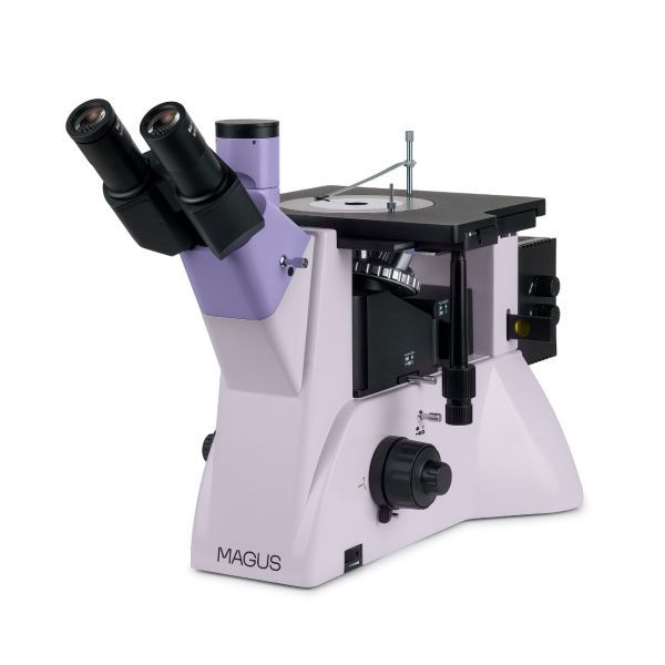
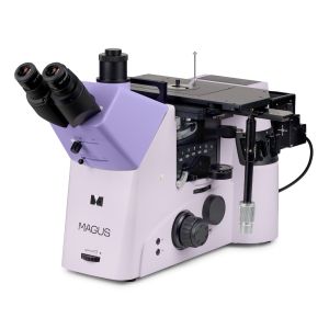
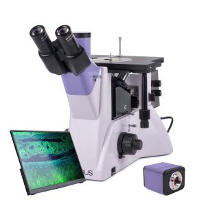
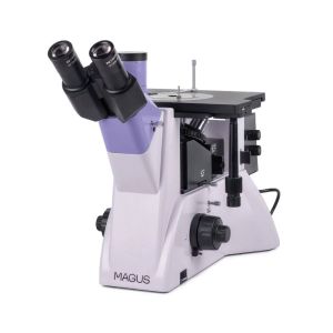
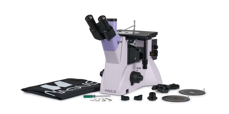
MAGUS Metal V700 DIC Metallurgical Inverted Microscope
Key features:
The microscope is designed for studying the microstructure of metals and alloys, semiconductor materials, and other opaque specimens.
The design of the inverted microscope does not limit the size of the examined sample; only its weight is limited up to 2kg. The specimen must be first specially processed, and then placed on the stage with the surface down.
The microscope implements three contrast techniques in reflected light: brightfield, simple polarization, and differential interference contrast. The brightfield method provides an authentic magnified image of the object. Simple polarization removes glare from metal surfaces and it highlights polymers and other foreign elements. Differential interference contrast colors the height difference of an object and thereby reveals the topography of the sample. The object of study will appear in relief.
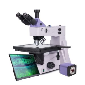
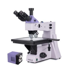
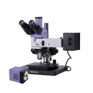
The trinocular tube and a camera side port enable the simultaneous connection of a monitor and camera.
The microscope can be used in various fields (metallurgical, engineering, aerospace, nuclear, and energy industries) as well as in research laboratories and technical universities.
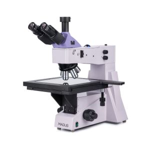
Microscope head
Tube length – infinity (∞). When assembling the microscope, the user can rotate the eyepiece tubes 180° and adjust the eye relief to fit their height. There is a trinocular head.
The monitor is mounted in the trinocular tube of the trinocular head and the digital camera – in the camera side port. The light beam on the trinocular head is split 50/50 (tube is open) or 100/0 (tube is closed), on the body – 100/0 or 0/100.
Revolving nosepiece
The 5-objectives revolving nosepiece is mounted on the stand under the stage.
The revolving nosepiece has a special DIC slider slot that is used when working in the differential interference contrast method with 5x, 10x, and 20x objectives.
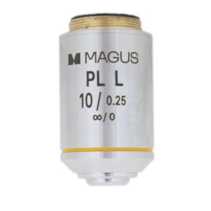
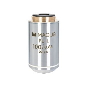
Objectives
Plan achromatic objectives with a long working distance that are designed for the brightfield, simple polarization, and DIC methods.
For the darkfield microscopy technique, choose the MAGUS Metal V700 BD.
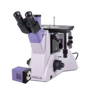
Focusing mechanism
Coaxial coarse and fine focusing knobs are located at the base of the microscope on both sides. The user can place their hands on the table and take a relaxed pose while observing. The focus adjustment is smooth and effortless.
On the right, there is the coarse focusing lock knob for quick adjustments after changing the sample. On the left side, there is the focusing tension adjusting ring.
Mechanical stage
The object is moved by moving the stage along two axes. A round rotating stage plate with a hole of suitable diameter (10mm, 20mm, or 30mm) is installed in the center of the stage. A specimen holder is used to hold the object in place.
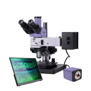
Light source
The lamphouse has a 50W halogen bulb. It is bright enough to observe with 4x to 100x objectives that are used for brightfield, polarized light, and DIC microscopy. The halogen bulb emits light with a color temperature that allows for comfortable work.
Reflected light illumination
The illumination system makes it possible to set up the Köhler illumination. The field and aperture diaphragms are pre-centered at the factory and require no additional centering. If necessary, the diaphragms can be adjusted with centering screws. The light source is centered along three axes.
The removable analyzer and polarizer are for the polarization microscopy technique. The polarizer rotates 0–360°, but the analyzer does not rotate.
The DIC prism slider is used when working by way of the differential interference contrast method with 5x, 10x, and 20x objectives.
A set of filters can help you adjust the color reproduction.
Accessories
Additional accessories increase the technical performance of the microscope. Eyepieces and objectives extend the range of magnification. Digital cameras output the image to a computer screen or monitor. Calibration slides help you take measurements of objects.
Specifications:
In the box:
Available on request:
All orders we receive until 15:00 concerning products in our warehouse, processed on the same day
Well-trained scientific and technical personnel are available to you at any time, to solve the questions and concerns about your equipment.
If you are not satisfied with the product you received, you can return it within 14 days of receipt.
Experience you can trust. Planetarium of Thessaloniki is the largest supplier of astronomy goods in the Balkans
 Ελληνικά