Language
 English
English
English
MAGUS
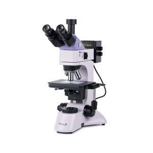
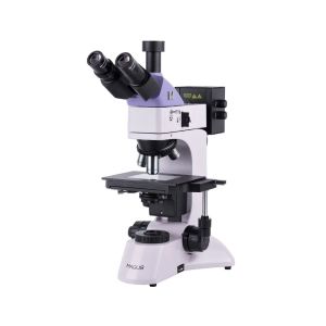
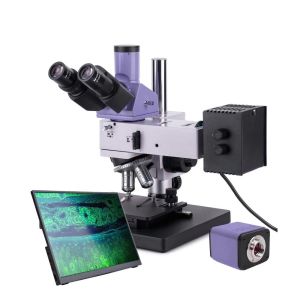
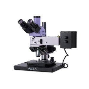
The microscope is designed for studying the microstructure of metals, alloys, semiconductor materials, paint coatings, and other opaque flat and polished samples. The reflected light illuminator allows for brightfield, darkfield, and polarization microscopy techniques.
Available Upon Request

This includes products that we do not keep in stock but are only available upon request or have been announced in advance by the manufacturer. These items are usually shipped within 5-14 days. Once we receive your order, we will notify you by e-mail or telephone of the exact delivery time, as well as the amount of any deposit that will be required.
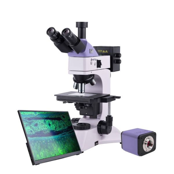
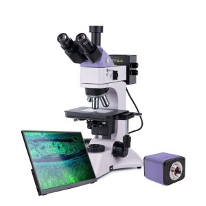
MAGUS METAL D600 BD LCD
Key features of the microscope:
Key features of the camera:
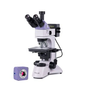
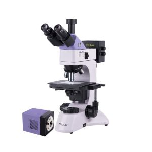
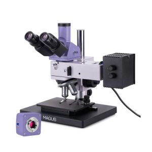
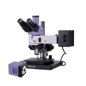
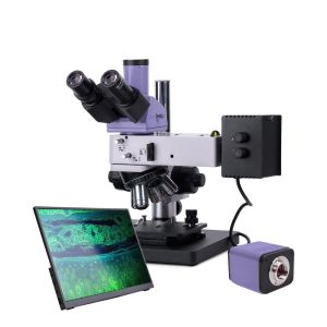
Digital camera
MAGUS CHD40 is a digital HDMI camera with three video interfaces and automatic switching between 4K and Full HD depending on the monitor resolution.
The camera is equipped with an 8MP sensor and produces realistic images in 4K resolution (3840x2160px) when connected via HDMI or USB3.0. When connected via Wi-Fi, the image quality is Full HD (1920x1080px).
The camera uses an HDMI interface to connect to a TV, monitor, or projector directly. In this mode, the camera operates autonomously without a connection to a PC. The HDMI interface provides a high and stable transfer rate from the camera to the external screen. You can connect the camera to a PC via Wi-Fi or USB3.0. Video is recorded at 30fps.
The camera combines high FPS and high bandwidth HDMI. Therefore, videos are vivid, with no freezes or gaps between frames. At maximum resolution, the image is well-detailed, moving objects are visible without bugs, and object movement is displayed without delays.
Monitor
The MAGUS MCD40 Monitor is designed to use a visualization system of the MAGUS microscope.
It is connected to a camera mounted on the microscope to display real-time images. It supports MAGUS 4K HDMI cameras.
The screen diagonal is 13.3 inches. The IPS matrix provides bright images with large viewing angles. If you look at the display at an angle, the color reproduction is not distorted.
The display can be placed on a folding stand on a table or shelf or mounted directly to the camera or microscope stand.
Microscope head
Trinocular head with infinity plan achromatic objectives. The digital camera is mounted in the trinocular tube. The eyepiece/camera light path selection knob allows you to direct the light beam to either the digital camera or the eyepiece tube. The diopter adjustment is on the left tube.
Revolving nosepiece
5 objectives. A free slot is used for centering the reflected light source. An additional objective can also be installed in the free slot to achieve extra magnification. The revolving nosepiece with objectives is oriented toward the interior – the user can see the objective inserted into the optical path, and the space above the stage is free.
Objectives
Plan achromatic objectives with long working distance designed for the brightfield and darkfield microscopy technique.
Focusing mechanism
Coaxial coarse and fine focusing knobs are located at the base of the microscope on both sides. The user can place their hands on the table and take a relaxed pose while observing. The focus adjustment is smooth and easy. On the right side, there is the coarse focusing lock knob and, on the left, there is a coarse focusing tension adjusting ring.
Stage
The object is moved by moving the stage along two axes. The maximum height of the observed sample must not exceed 20mm. The glass stage plate is used for observing translucent specimens.
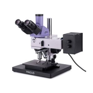
Light source
Halogen bulbs are used for both reflected and transmitted illumination. Halogen bulbs emit light with a color temperature that allows for comfortable work. The 30W bulb is bright enough to observe with 4x to 100x magnification objectives that are used for brightfield and polarized light microscopy. The reflected light illuminator has a 50W bulb for comfortable darkfield observations.
Reflected light illumination
The microscope is equipped with a built-in analyzer and a removable polarizer. The polarizer rotates 0–360°, the analyzer does not rotate. The aperture and field diaphragms make it possible to set up the Köhler illumination. Both diaphragms and the light source can be centered. The microscope comes with a set of filters. The darkfield device is integrated into the reflected light illuminator.
Transmitted light illumination
An adjustable field diaphragm, a center-adjustable and height-adjustable Abbe condenser with NA 1.25 allow to set up the Köhler illumination. The condenser has a flip-down lens for low magnification objectives.
Accessories
There is a broad range of accessories to be used with the microscope: eyepieces, objectives, digital cameras, calibration slides, etc.
Microscope specifications
Camera specifications
Monitor specifications
In the box:
Available on request:
All orders we receive until 15:00 concerning products in our warehouse, processed on the same day
Well-trained scientific and technical personnel are available to you at any time, to solve the questions and concerns about your equipment.
If you are not satisfied with the product you received, you can return it within 14 days of receipt.
Experience you can trust. Planetarium of Thessaloniki is the largest supplier of astronomy goods in the Balkans
 Ελληνικά