Language
 English
English
English
MAGUS
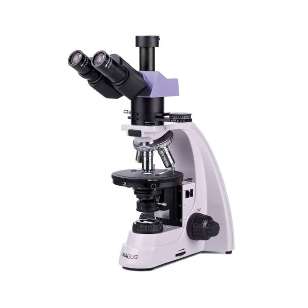
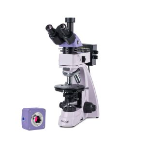
The microscope is designed for studying anisotropic geological, biological, and polymeric samples in polarized and transmitted light.
Available Upon Request

This includes products that we do not keep in stock but are only available upon request or have been announced in advance by the manufacturer. These items are usually shipped within 5-14 days. Once we receive your order, we will notify you by e-mail or telephone of the exact delivery time, as well as the amount of any deposit that will be required.
MAGUS POL 800
Key features
Microscope head
Trinocular head with infinity plan achromatic objectives. Digital image capturing devices are mounted in the trinocular tube. The eyepiece/camera light path selection knob allows you to direct the light beam to either the digital camera or the eyepiece tubes.
The diopter adjustment is on the left tube.
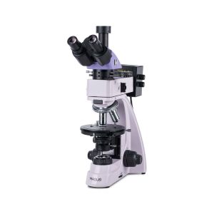
Revolving nosepiece
Five objectives. An additional objective can also be installed in a free slot in order to achieve extra magnification. The design of the revolving nosepiece (“away from the observer”) frees up space at the front of the stage so that the user can see the objective inserted into the optical path. The revolving nosepiece slots are centered to align the optical axis of the objective and microscope.
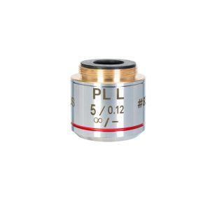
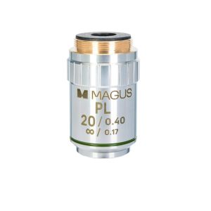
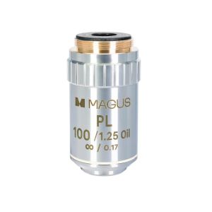
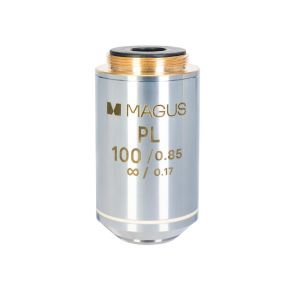
Objectives
Infinity plan achromatic objectives are designed specifically for polarized light observations: The strain-free optics ensure that the birefringence comes from the specimen and not from the optical elements. Designed for use with 0.17mm thick cover slips.
Focusing mechanism
Coaxial coarse and fine focusing knobs are located at the bottom of the stand on both sides. The user can place their hands on the table and take a relaxed pose while observing. The focusing adjustment is smooth and effortless.
The design of the focusing mechanism provides for quick microscope adjustment after changing the object of study. For this purpose, a coarse focusing lock knob is located on the right side. The coarse tension can be adjusted by turning the ring on the left side.
Stage
The stage rotates 360° to view the color change of the sample when the polarizer and analyzer are in crossed orientation. The stage has a gradation of the rotation angle. With the vernier scale, measurements are made with an accuracy of 0.1°.
The stage can be centered with two screws because the analysis of an anisotropic object in polarized light requires the precise alignment of the rotation axis of the stage with the optical axis of the microscope.
Light source
Halogen bulbs emit light with a color temperature that allows for comfortable work. The 30W bulb is bright enough for you to observe with the 4x to 100x magnification objectives that are used for brightfield and polarized light microscopy.
Transmitted light illumination
Köhler illumination: adjustable field diaphragm; centered and height-adjustable Abbe condenser with adjustable aperture diaphragm and swivel auxiliary lens. NA = 1.25. The polarizer is 0–360° rotatable, with four rotation angles of 0°, 90°, 180°, 270° marks on the scale. The analyzer rotates 90° and the vernier scale on the analyzer provides accurate angle measurement.
Köhler illumination
The Köhler illumination setup enhances the image quality of a specimen. With such illumination, you can achieve maximum resolution on each objective and the uniform illumination of the field of view with no darkening at the edges. The object of study is in sharp focus and the image artifacts are removed.
Studying samples in polarized light
The intermediate tube holds the analyzer and Bertrand lens, and it has a slot for compensators. The analyzer is inserted into the light path to observe objects in polarized light. When the polarizer and analyzer rotate, the polarization angle changes. You can achieve a mutually perpendicular position by setting both filters to the “0” position. When the microscope stand is rotated, depending on the polarization angle, the surface of the sample reflects light differently. The Bertrand lens is used for conoscopic studies. The compensators are designed to enhance the contrast of samples with weak birefringence.
Accessories
There is a line of accessories designed specifically for this microscope.
Additional objectives can be used for studying objects up to 0.22µm.
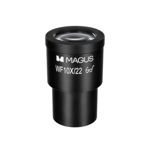
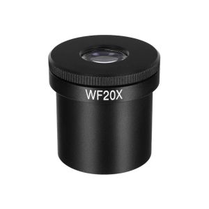
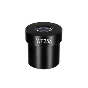
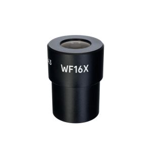
Eyepieces extend the magnification range of the microscope. Additional eyepieces will help you to utilize the full potential of an objective that is used more often.
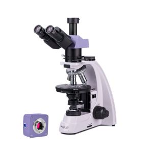
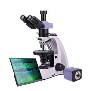
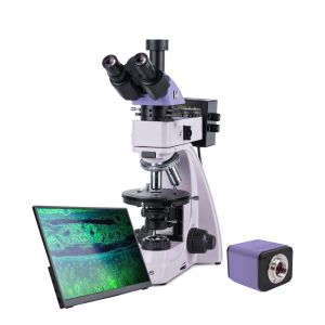
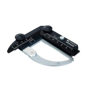
A digital camera that outputs the microscope image on a monitor, stores files, and that has software that takes real-time measurements of specimens. A calibration slide for measuring objects that can be combined with an eyepiece with a scale or the camera software.
Specifications
In the box:
Available on request:
All orders we receive until 15:00 concerning products in our warehouse, processed on the same day
Well-trained scientific and technical personnel are available to you at any time, to solve the questions and concerns about your equipment.
If you are not satisfied with the product you received, you can return it within 14 days of receipt.
Experience you can trust. Planetarium of Thessaloniki is the largest supplier of astronomy goods in the Balkans
 Ελληνικά