Language
 English
English
English
MAGUS
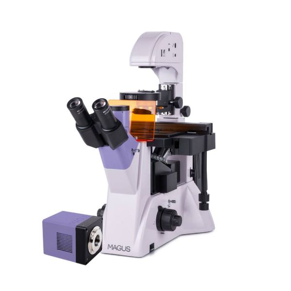
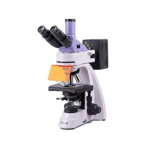
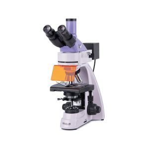
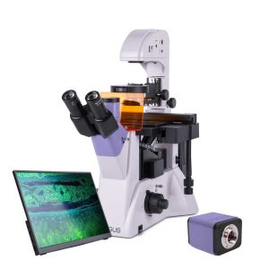
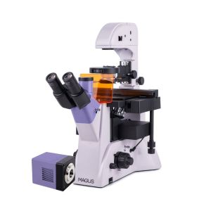
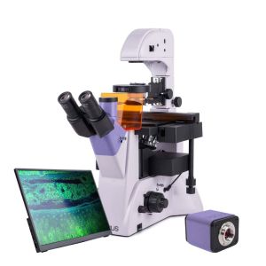
The microscope is intended for studying specimens using the fluorescence microscopy technique in reflected light and using the brightfield and phase contrast techniques in transmitted light.
Available Upon Request

This includes products that we do not keep in stock but are only available upon request or have been announced in advance by the manufacturer. These items are usually shipped within 5-14 days. Once we receive your order, we will notify you by e-mail or telephone of the exact delivery time, as well as the amount of any deposit that will be required.
MAGUS LUM VD500
Key features of the microscope:
Key features of the camera:
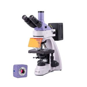
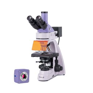
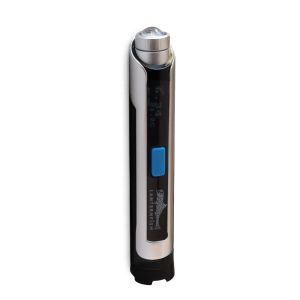
Digital camera
The MAGUS CLM90 digital camera is designed for use in fluorescence and darkfield microscopy techniques.
The camera is equipped with a 7.1MP sensor and produces realistic images at a resolution of 3200x2200px. The camera is recommended for use with 4x, 10x, 20x, and 40x objectives. When working with low magnification objectives, the camera will allow you to see the finer details.
Video is recorded at 51.3fps or 133.8fps depending on its resolution. Videos are smooth with soft and subtle transitions between frames. The movement of the sample is displayed in real time with no delays. The camera makes it easy to work with objects that are moving and is suitable for classroom demonstrations.
The camera is equipped with a USB3.0 interface. The data transfer speed is 10 times faster than USB2.0 cameras. The high-speed camera is recommended for professional laboratories, research, or university training.
Microscope head
Tube length – infinity (∞). When assembling the microscope, the user can rotate the eyepiece tubes 180° and adjust eye relief to fit their height. There is a trinocular microscope head.
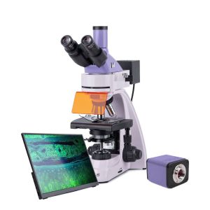
The monitor is mounted in the vertical tube of the trinocular head and the digital camera – in the camera side port. The trinocular head has an 80:20 beam splitter, and there is a 2-position beam splitter on the body (100:0/0:100).
Revolving nosepiece
The 6-objectives revolving nosepiece is mounted on a stand under the stage.
Objectives
Infinity plan achromatic objectives with long working distance for dishes with a bottom thickness of 1.2mm.
Three objectives are intended for brightfield and fluorescence microscopy techniques and the other 3 objectives are for the phase contrast technique.
Focusing mechanism
Coaxial coarse and fine focusing knobs are located at the base of the microscope on both sides. The user can place their hands on the table and take a relaxed pose while observing. The focusing adjustment is smooth and effortless. On the right, there is a coarse focusing lock knob for quick adjustments after changing the specimen. On the left, there is a coarse tension adjustment ring for further adjustment.
Stage
There is a fixed stage with a mechanical attachment for moving the laboratory ware in two mutually perpendicular directions. The smooth and subtle movement of the object augments the accuracy of the study – no part of the specimen will be overlooked. The travel knob is located at the bottom and so the user will not strain their right hand. The microscope set includes four dish holders for dishes of different sizes.
Reflected light illuminator
The fluorescence excitation source is a 100W mercury lamp. It is extremely bright and has a wide spectrum of wavelengths. The mercury lamp has discrete peaks, allowing you to work with many fluorochromes.
The reflected light illuminator contains four excitation filters: ultraviolet (UV), violet (V), blue (B), and green (G).
The mercury lamp is located in the lamphouse and can be centered in three planes. The heat radiation is conducted away from the lamphouse and, therefore, the lamp does not get too hot. The mount allows for safe and quick lamp replacement.
Condenser
Phase contrast turret condenser with four positions. There is a free slot for brightfield microscopy; the other three slots with phase annuli plates are used for working with objectives at magnifications of 10x, 20x, and 40x. The phase contrast rings can be centered. Fast and intuitive switching between microscopy techniques saves time and simplifies the researcher’s work.
Reflected light source
The reflected light illuminator has a 30W halogen bulb. Halogen bulbs emit light with a color temperature that allows for comfortable work. The 30W bulb is bright enough to observe with all the objectives that are used for brightfield and phase contrast microscopy techniques.
Köhler illumination in transmitted light
The Köhler illuminator improves the image quality of the observed sample: Each objective achieves maximum resolution. The field of view is illuminated evenly without darkening at the edges. The object of study is in sharp focus and the image artifacts are removed.
Accessories
There is a line of accessories designed specifically for this microscope.
Eyepieces that extend the magnification range of the microscope. The optional eyepieces help you maximize the potential of the objective that you use most often.
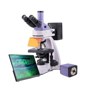
A digital camera that outputs the microscope image on a monitor and stores files along with software that takes real-time measurements of specimens.
A calibration slide for measuring objects that can be combined with an eyepiece with a scale or the camera software.
Microscope specifications
Camera specifications
In the box:
Available on request:
All orders we receive until 15:00 concerning products in our warehouse, processed on the same day
Well-trained scientific and technical personnel are available to you at any time, to solve the questions and concerns about your equipment.
If you are not satisfied with the product you received, you can return it within 14 days of receipt.
Experience you can trust. Planetarium of Thessaloniki is the largest supplier of astronomy goods in the Balkans
 Ελληνικά