Language
 English
English
English
MAGUS
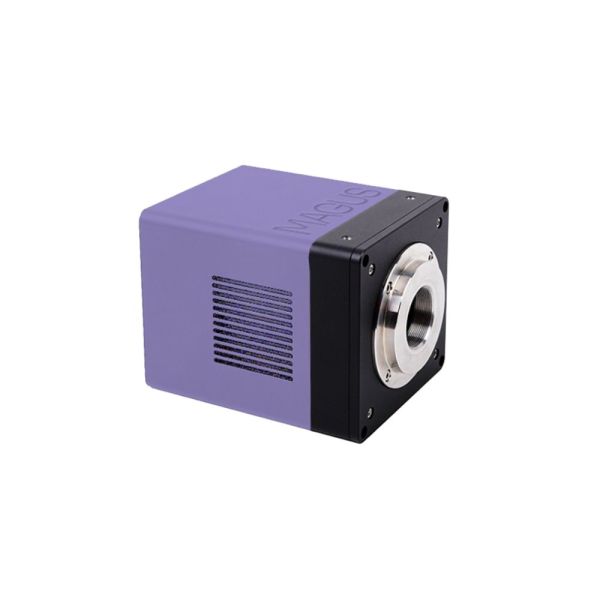
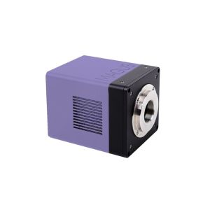
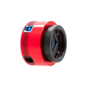
The MAGUS CLM30 digital camera is designed for use in fluorescence and darkfield microscopy techniques. The high-speed camera is recommended for professional laboratories, research, or university training.
Available within 5-14 days

This includes products that we do not keep in stock but are only available on request or have been announced by the manufacturer. For orders of these items, a deposit is required, proportional to the total value of your order. As soon as we receive your order, we will notify you by e-mail or telephone, for the exact delivery time and the amount of the deposit.

MAGUS CLM30 DIGITAL CAMERA
Features:
Sensor
The camera is equipped with a SONY Exmor backlit color CMOS sensor. This technology increases light sensitivity and improves camera performance in low-light conditions.
A color sensor has less light sensitivity than a monochrome one, but it is absolutely necessary for studies that identify and classify objects based on color.
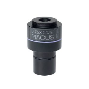
The 1/1.2" sensor with 2.9x2.9µm pixel provides light sensitivity of 5970mV with 1/30s. To achieve the maximum field of view without distortion, we recommend using an adapter with a magnification of 0.75x to 1x.
Peltier element
The camera sensor may overheat during prolonged use. This causes thermal noise in the image, which significantly reduces the image quality. To eliminate this negative effect, the camera is equipped with a two-stage thermoelectric module (Peltier element). It sets the temperature to 42°C below room temperature and creates optimal conditions for the sensor to run for long periods of time without heating up. Therefore, the camera is suitable for long shutter speeds: The image is clear without thermal noise.
Software features
The camera uses software to display images on an external display, take photographs, record video, edit images, and measure linear and angular dimensions of samples and their structures. Before taking measurements, the software must be calibrated for each objective with a calibration slide.
Installation
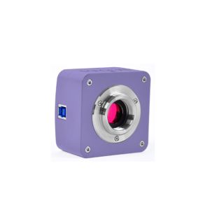
The camera is mounted on the microscope in the trinocular tube/camera port using the C-mount adapter (included).
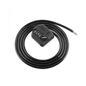
In the box:
All orders we receive until 15:00 concerning products in our warehouse, processed on the same day
Well-trained scientific and technical personnel are available to you at any time, to solve the questions and concerns about your equipment.
If you are not satisfied with the product you received, you can return it within 14 days of receipt.
Experience you can trust. Planetarium of Thessaloniki is the largest supplier of astronomy goods in the Balkans
 Ελληνικά